Unveiling integrin beta-6 prevalence in NSCLC with real-world data
RWD analysis shows high IB6 expression in nonsquamous mNSCLC
Molecular characteristics by IB6 IHC status
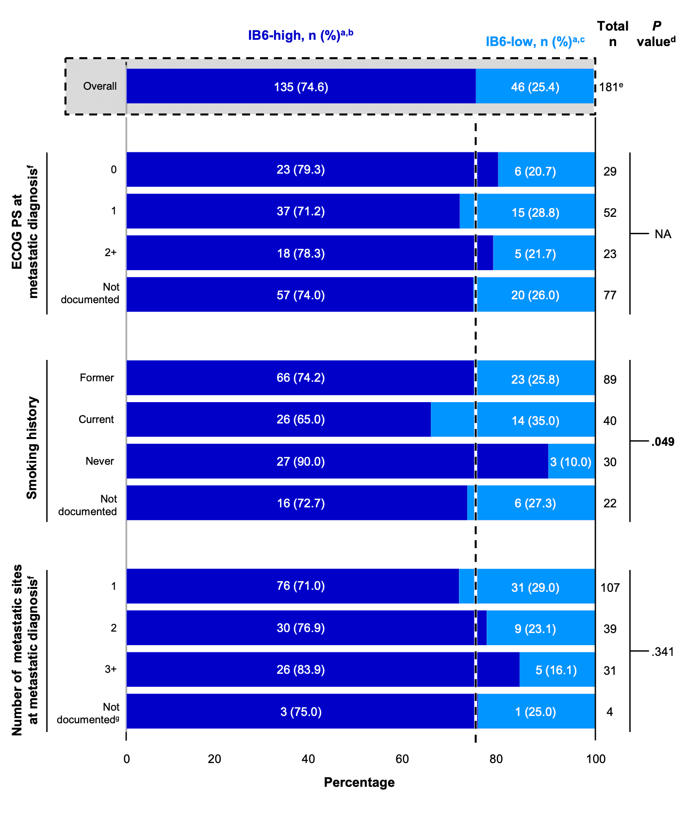
PD-L1, programmed cell death ligand 1; TPS, tumor proportion score. a Percentage was calculated with “total n” as denominator for each row. b IB6-high expression was defined as ≥50% positivity of tumor cells at ≥2+ intensity. c IB6-low expressors was defined as <50% positivity of tumor cells at ≥2+ intensity. d Comparisons were not made for characteristics with >20% missing data. P values were calculated using Fisher exact test. e Includes only evaluable patients with a known IB6 IHC status. The other 19 patients had failure at the hematoxylin & eosin evaluation by PathAI due to insufficient tumor or fine needle aspiration cell blocks. f ALK fusions were determined using DNA, a combination of DNA and RNA if DNA evidence was insufficient, or RNA only. g__Includes the following EGFR mutations: classic exon 19 deletion, L858R, exon 20 Insertion, exon 19 in-frame deletion, exon 19 deletion-insertion, exon 19 in-frame insertion, exon 20 substitution, and exon 4 mutation..
 Lens Library

