Indication Lung

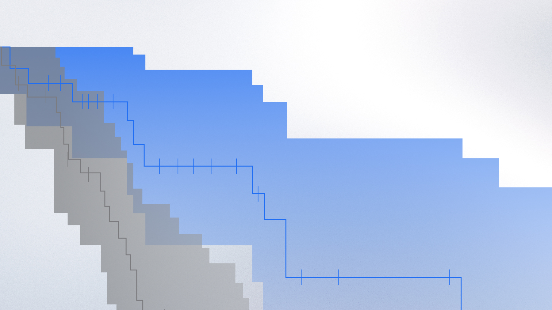
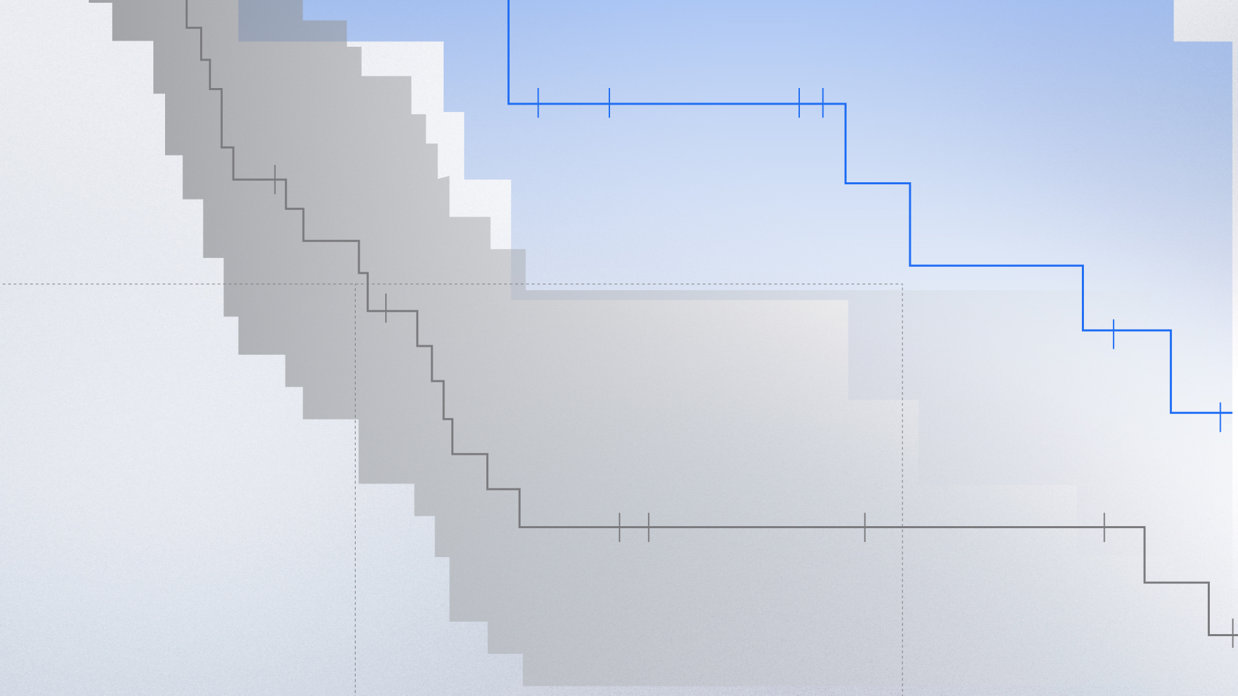
Characterizing the immune microenvironment across NSCLC metastatic sites

RWD analysis reveals distinct immune profiles between primary and metastatic NSCLC tumors

Indication Lung

PD-L1 positive status was lower in liver, CNS, and bone compared to lung (***q < 0.001, **q < 0.01)

Introduction and Hypothesis

The efficacy of immune checkpoint inhibitors (ICIs) in non-small cell lung cancer (NSCLC) is heavily influenced by the tumor immune microenvironment (TIME). However, the TIME is often characterized using primary tumor biopsies, which may not reflect the biology of metastatic sites where disease progression occurs. Researchers hypothesized that the TIME of common metastatic sites—such as the liver, central nervous system (CNS), and bone—is immunologically distinct from that of the primary lung tumor. Understanding these differences is critical for optimizing treatment strategies and informing the design of future clinical trials. The research team chose to leverage a large, multimodal real-world database to investigate these site-specific differences at a scale not achievable through traditional clinical studies.

Methodology

Researchers utilized Tempus Lens to conduct a retrospective analysis of a de-identified cohort of 6,534 patients with metastatic NSCLC from a multimodal real-world database. The analysis integrated solid-tumor DNA sequencing from a 648-gene panel (Tempus xT) with whole-transcriptome RNA sequencing (Tempus xR). A key analytical feature was an immune infiltration algorithm that deconvolved RNA expression data to estimate the proportions of various immune cell types within a sample. The patient cohort was stratified based on the anatomical site of the tumor biopsy (primary lung, liver, CNS, or bone) to compare the TIME and key immuno-oncology biomarkers, including immune cell infiltration, PD-L1 expression and tumor mutational burden (TMB), across these distinct groups.

Impact

The analysis revealed that metastatic sites in the liver, CNS, and bone are significantly less immunogenic, or immunologically “colder,” than primary lung tumors. These metastatic tumors showed significantly lower infiltration of key anti-tumor immune cells, including CD4+ and CD8+ T cells (q<0.001), while showing a higher percentage of macrophages. Correspondingly, PD-L1 positive status was lower in metastatic sites compared to the lung. 

In a notable finding, CNS metastases had a significantly higher frequency of TMB-High status compared to lung tumors (q<0.001). For life sciences companies, this research may provide critical evidence that the primary tumor’s immune profile is not always representative of that of metastatic disease. These insights can directly inform the development of novel therapies and help refine clinical trial designs to account for site-specific immune composition, ultimately improving the efficacy of immunotherapies for patients with metastatic NSCLC.

LEARN MORE

Contact us

Learn more about Tempus or explore more Lens research projects.