-
PROVIDERS
REGISTER NOW
Upcoming Webinar:
MRD data insights across the cancer care continuum -
LIFE SCIENCES
REGISTER NOW
From insight to impact: Leveraging the AI-enabled Next platform with BMS to advance equitable access in precision oncology
-
PATIENTS
It's About Time
View the Tempus vision.
- RESOURCES
-
ABOUT US
View Job Postings
We’re looking for people who can change the world.
- INVESTORS
RADIOLOGY /// CARDIO
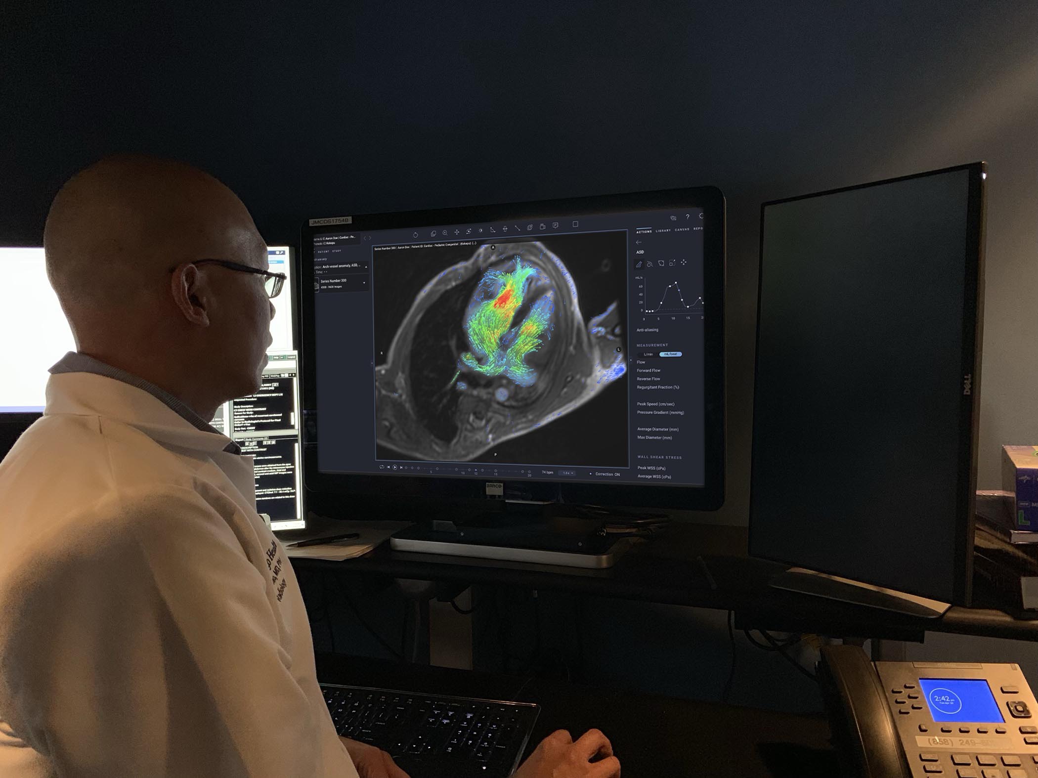
AI-enabled solution to analyze and quantify cardiac MR images1
Tempus Pixel provides advanced viewing and automated reporting of cardiac MR images to help improve efficiency and accuracy in flow visualization and quantification, functional analysis, and tissue characterization.
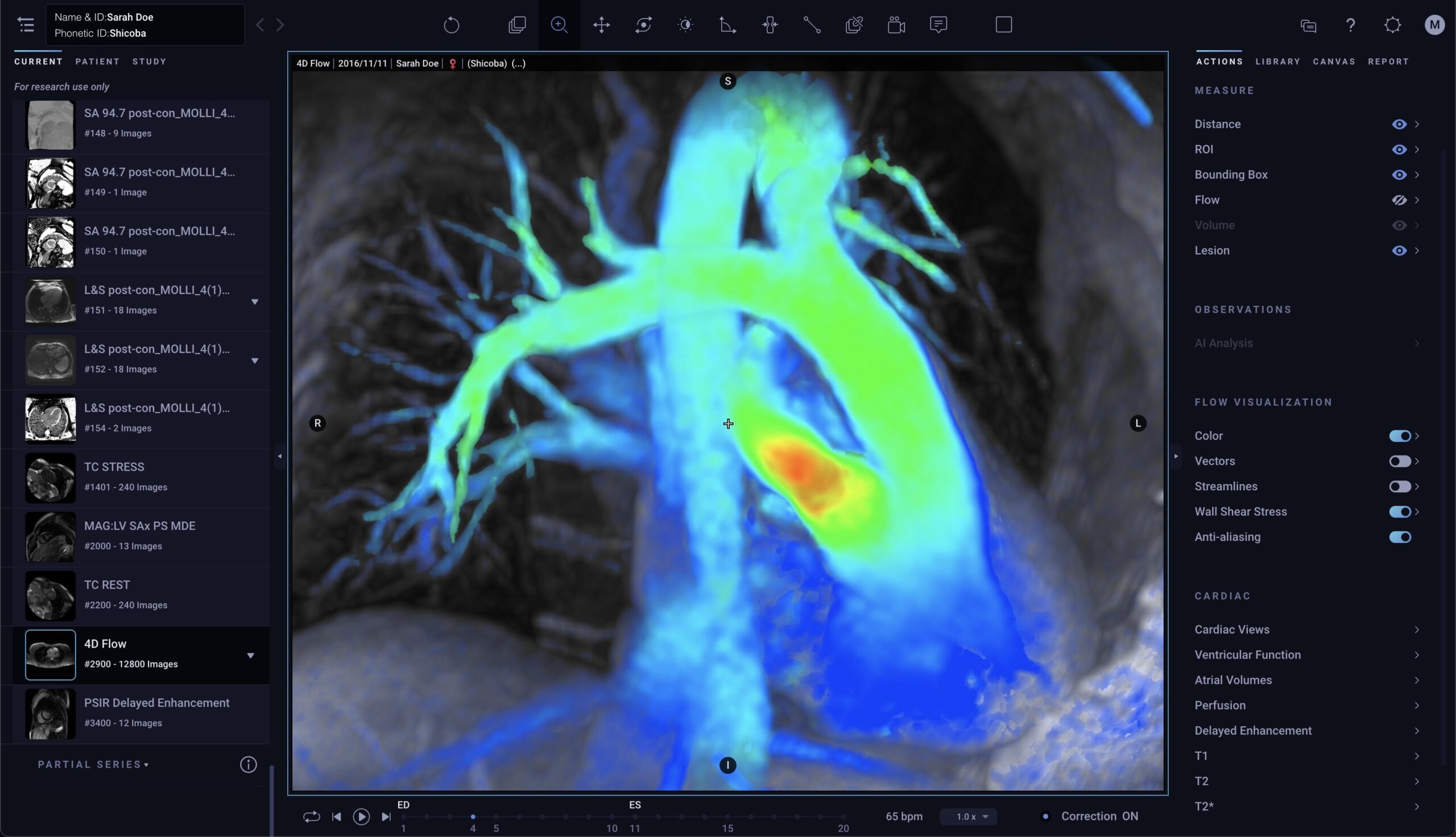
AI-enabled 4d flow
Visualize and quantify blood flow precisely anywhere in the heart and its major vessels based on 4D flow datasets, enabling reductions in scan time of up to 30%2
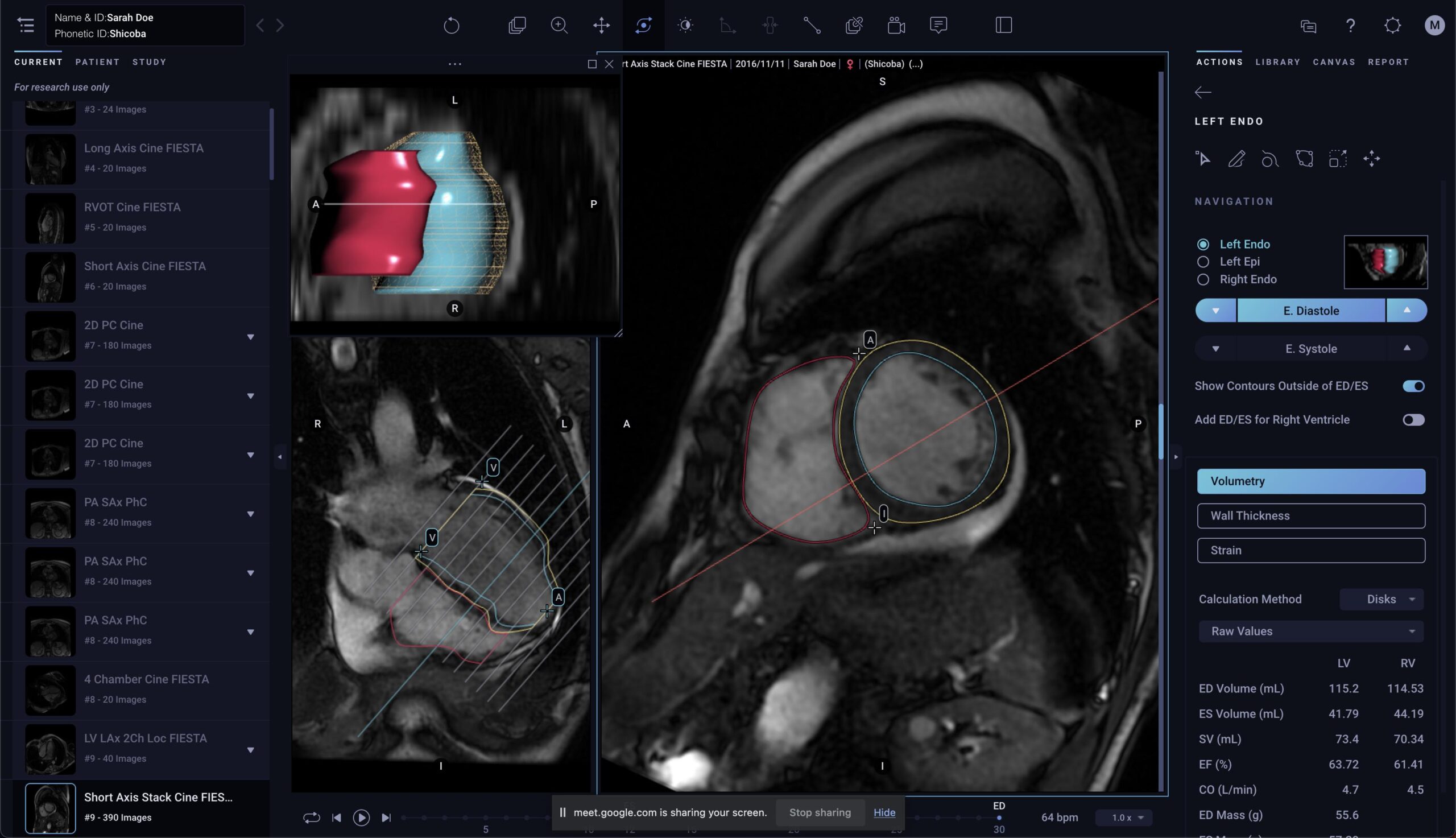
functional analysis
Automatically quantify cardiac chambers volumetry and ventricular function data from short axis datasets, enabling a reduction in segmentation time of up to 93% per study3,4
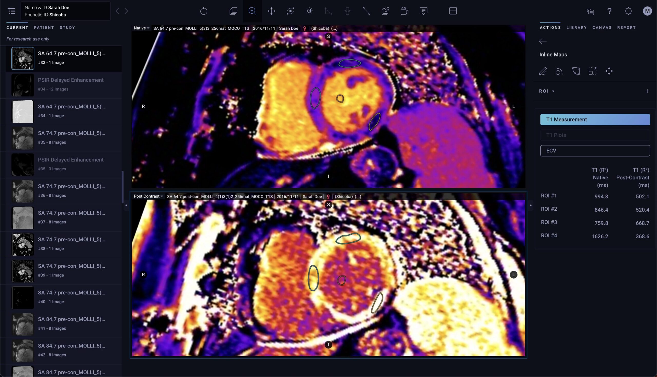
tissue characterization
Automatically quantify delayed enhancement, perfusion, and parametric mapping images (T1, T2 and T2*), with graphs and 17-segment models for faster and more informed decisions5
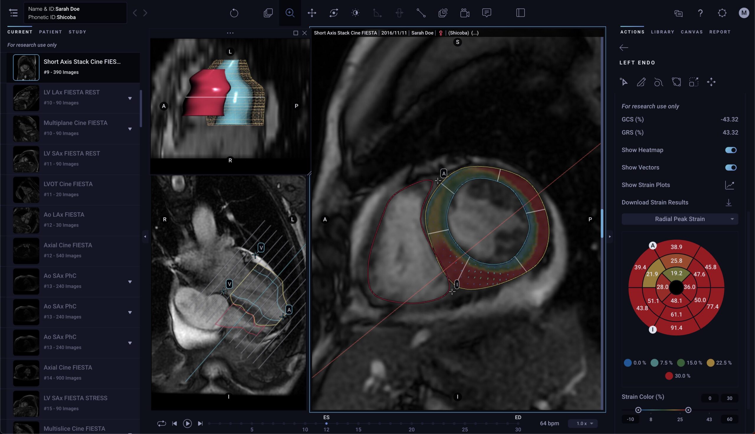
AI-based Myocardial Strain
Calculates strain, strain rate, and myocardial velocity that are reliable, quantitative, visually confirmable, and that separates normal from disease myocardial tissue6
seamless integration
Easily integrates AI-enabled tools with existing PACS, EHR, worklist, notification, and dictation systems to optimize radiology workflows
-
UPCOMING WEBINAR:
Radiology: Cardiothoracic Imaging
4D Flow MRI Quantification of Congenital Shunts: Comparison to Invasive Catheterization
Read more -
UPCOMING WEBINAR:
JACC: Cardiovascular
AI Based CMR Assessment of Biventricular Function: Clinical Significance of Intervendor Variability and Measurement Errors
Read more -
UPCOMING WEBINAR:
BMC Medical Imaging
Quantitative evaluation of aortic valve regurgitation in 4D flow cardiac magnetic resonance: at which level should we measure?
Read more -
UPCOMING WEBINAR:
Radiology: Cardiothoracic Imaging
Deep Learning Synthetic Strain: Quantitative Assessment of Regional Myocardial Wall Motion at MRI
Read more -
UPCOMING WEBINAR:
BMC Medical Imaging
Right and left ventricular function and flow quantification in pediatric patients with repaired tetralogy of Fallot using four-dimensional flow magnetic resonance imaging
Read more -
UPCOMING WEBINAR:
The American Journal of Cardiology
Relation of Myocardial Perfusion Reserve and Left Ventricular Ejection Fraction in Ischemic and Nonischemic Cardiomyopathy
Read more
-
Tempus Pixel Cardio is FDA-cleared (K203744) and CE Marked. Arterys Inc is the manufacturer of Tempus Pixel Cardio.
- Atkins MB. Integration of 4D-Flow into routine clinical practice of congenital and non-congenital cardiac MRI-18 months experience demonstrating decreased scan times, physician monitoring, and patient breath hold times. [366897]. CMR 2018; pp. 789-790.
- Narang A, Miller T, Ameyaw K, et al. A machine learning algorithm fordetermination of left ventricle volumes and function: comparison to manual quantification. J Am Coll Cardiol. 2019;73(9):1638. https://www.researchgate.net/publication/331516422_A_MACHINE_LEARNING_ALGORITHM_FOR_DETERMINATION_OF_LEFT_VENTRICLE_VOLUMES_AND_FUNCTION_COMPARISON_TO_MANUAL_QUANTIFICATION
- Retson T, Masutani E, Chen C, et al. Real-world clinical performance of deep learning for quantification and segmentation of biventricular cardiac size and function [4785], Proc Intl Soc Mag Reson Med. 2018;26 4785. https://cds.ismrm.org/protected/18MProceedings/PDFfiles/4785.html
- T2* Parametric mapping is only supported for GE and Siemens scanners.
- Strain is for Research Use Only in the EU and US.