-
PROVIDERS
New MRD Medicare Coverage for Select Indications*
*When coverage criteria are met. Additional criteria and exceptions for coverage may apply.
-
LIFE SCIENCES
Register now
UPCOMING WEBINAR
Unlocking Foundation Models: Our experience from proof of concept to deploying at scale -
PATIENTS
It's About Time
View the Tempus vision.
- RESOURCES
-
ABOUT US
View Job Postings
We’re looking for people who can change the world.
- INVESTORS
02/03/2026
Spatial Transcriptomics Vendor Solutions: Tempus Technical Platforms and Capabilities
Main Takeaways:
- Tempus offers the 10x Visium platforms for spatial transcriptomics.
- 10X Genomics Visium HD platform which achieves whole-transcriptome profiling at 2 𝜇m resolution.
- Tempus supports clinical development from biomarker discovery to companion diagnostic development as an end-to-end provider.
Omics Solutions at Tempus
Tempus offers a wide range of disease-agnostic technologies, including NGS, methylation testing, genotyping, proteomics, and single-cell sequencing, designed to support all phases from biomarker discovery to clinical development. Our research use only (RUO) omics testing solutions, alongside our vast data repository, biological modeling solutions, and sequencing tests, can offer comprehensive insights for disease mechanism analysis and translational research. Tempus supports clinical development from biomarker discovery to companion diagnostic development as an end-to-end provider. Tempus offers the 10x Visium platforms for spatial transcriptomics. Utilize the 10x Visium V2 at 25K read pairs per tissue-covered spot or Visium HD for 275M read pairs for fully captured area. Streamlined Vendor Management With solutions throughout the clinical development and research process, Tempus can be your sole vendor. White Glove Project Management Receive real-time project updates from a dedicated project manager throughout the project’s duration.
Visium HD combined with deep-learning-based cell segmentation on H&E images yield accurate cell annotation at single-cell resolution
NGS-based Spatial Transcriptomics (ST) technologies have gained increasing attention for their ability to provide spatial context of gene expression, but they have been constrained by low resolutions until the recent launch of 10X Genomics Visium HD platform which achieves whole-transcriptome profiling at 2 𝜇m resolution. Here we aim to solve that challenge by combining Visium HD data with custom deep-learning-based cell segmentations to yield accurate single-cell level data and enable downstream clustering and cell type annotations. We demonstrate the feasibility and advantages of analyzing Visium HD data at single-cell resolution using deep-learning-based segmentation models applied to H&E images. Cell clusters and LLM-based cell type annotations derived from single-cell resolution Visium HD data are highly consistent with pathologist annotations and morphology-based cell classification models.
Comparison of interassay similarity and cellular deconvolution in spatial transcriptomics data using Visum CytAssis
We used the 10X Visium CytAssist platform to generate ST data and additionally generated paired bulk RNAseq data. To test the interassay reliability of CytAssist on archival FFPE tissue sections, we compared ST results across 3 sample preparation conditions. Our findings demonstrate the feasibility and translational utility of ST to discover spatial signatures and the cellular context in retrospective clinical cohorts to empower discovery and translational efforts in precision oncology and therapeutic development.
Tempus Publication – Comparison of Interassay Similarity and Cellular Deconvolution in Spatial Transcriptomics Data Using Visium CytAssist
Spatial Transcriptomics (ST) is an emerging technology that characterizes gene expression within the spatial context of tissue. ST data can be generated directly from archival formalin fixed paraffin embedded samples, enabling the study of spatial gene expression in real-world clinical settings. To test interassay reliability of CytAssist on archival FFPE tissue sections, we selected FFPE tissue sections from 6 NSCLC and 1 tumor of unknown origin (TUO) samples from patients in the Tempus database. We find key quality control metrics and spatial gene expression patterns are consistent across 3 different H&E staining protocols (Figs 1,3,4). Specifically, our results show clinical archival FFPE samples yield high interassay reliability via the CytAssist platform.
Tempus Publication – Analysis of multimodal real world data from 1,248 breast cancer patients
We assembled and analyzed a multi-modal real-world cohort of 1,248 breast cancer patients, enriched for pre- and post-CDK4/6i, to derive a comprehensive molecular landscape of CDK4/6i resistance. Spatial transcriptomics data revealed HR+ tumors comprise of LumA, LumB and Her2 cells co-localized within tumor regions. These suggest that the expansion/emergence of aggressive subtype represents a major mechanism of CDK4/6i resistance.
The operating system for oncology R&D
Spatial biology techniques now provide high-resolution views of the tumor microenvironment (TME), allowing researchers to map the location of cells, transcripts, and proteins within intact tissues, revealing how spatial arrangement influences tumor progression, immune infiltration, and therapeutic response—insights that go far beyond the average signals of bulk molecular analysis. To further enhance discovery and characterization, Tempus also provides single-cell RNA sequencing and spatial transcriptomics to map cell populations and to better characterize the relationships between molecular profiles and therapeutic response. Tempus delivers an integrated platform of multimodal data, advanced analytics, and AI-driven applications to support the next era of precision oncology. Access one of the world’s largest oncology multimodal real-world databases, linking DNA, RNA, H&E images, and other clinical, imaging, and molecular data to uncover insights across the R&D continuum.
-
 03/27/2026
Impact of M2-like macrophage infiltration on RCC immunotherapy outcomes
Tempus Lens analysis reveals how immune profiling enhances macrophage predictive value
Read more -
03/27/2026
Developing a gene expression signature for first-line treatment in metastatic CRC
Tempus RWD identifies a signature predictive of bevacizumab response in CRC
Read more -
 03/27/2026
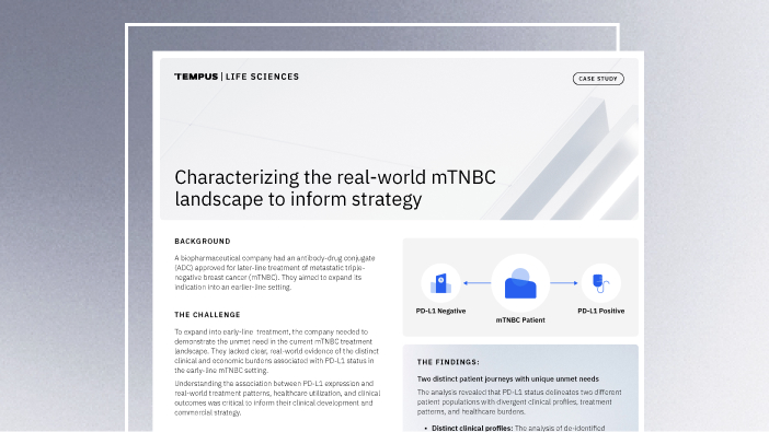
Characterizing the real-world mTNBC landscape to inform strategy
Learn how Tempus’ analysis of multimodal real-world data helped a biopharmaceutical company identify distinct patient journeys and healthcare burdens in mTNBC based on PD-L1 status, enabling them to refine their clinical and commercial strategy.
Read more