-
PROVIDERS
New MRD Medicare Coverage for Select Indications*
*When coverage criteria are met. Additional criteria and exceptions for coverage may apply.
-
LIFE SCIENCES
Register now
UPCOMING WEBINARTranslating data into an actionable R&D strategy
-
PATIENTS
It's About Time
View the Tempus vision.
- RESOURCES
-
ABOUT US
View Job Postings
We’re looking for people who can change the world.
- INVESTORS
01/05/2026
Multiomic Analysis and Oncologic Outcomes in Pancreatic Cancer by PIN1 Expression
ASCO GI 2026
PRESENTATION
Authors
Mustafa Raoof, Adam Dugan, Maurizio Pellecchia, Allison Rosenzweig, Loretta Erhunmwunsee, Stamatina Fragkogianni, Jacob Mercer, Ezra E.W. Cohen, Thatcher Heumann, Justin H. Lo
Background: Proline isomerase (PIN1) is a novel investigational target and is associated with desmoplastic stroma, an immunosuppressive tumor immune microenvironment (TIME) and worse outcomes in pancreatic ductal adenocarcinoma (PDAC). However, stromal targeting has shown conflicting evidence, with some studies showing it leads to more aggressive tumors. To further understand the implications of targeting PDAC stroma, we characterized PIN1 expression and its impact on the TIME and survival.
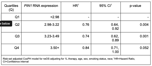
Methods: Patients with a primary diagnosis of PDAC and a stage 4 biopsy sequenced with Tempus xT DNA and/or xR whole transcriptome analysis prior to receiving either 1L FOLFIRINOX or gemcitabine + nab-paclitaxel were selected from the Tempus LENS deidentified database (n = 1844). Samples were from the liver (L; 49.7%), primary tumor (PT; 26.4%), peritoneum (PT; 12.4%), or non-liver or peritoneal sites (NLP; 11.6%). RNA-sequencing data were corrected for batch and assay effects and then normalized by computing log2(TPM+1) (TPM = transcripts per million). PDAC samples were stratified into quartiles (Q1-Q4; n = 461 for each) by PIN1 RNA expression. Immune cell proportions were estimated from RNA (quanTIseq). PD-L1 was assessed by IHC using the 22C3 clone. Cox proportional hazards (CoxPH) models with risk set adjustment were used to test for adjusted associations between PIN1 and real-world overall survival (rwOS). All p-values were uncorrected. Statistical significance was defined as p < 0.05.
Results: The PDAC cohort was composed of a diverse population: 56% White, 6.3% Black/African-American, 1.7% Asian and 36.2% Other/Unknown. PIN1 RNA expression was higher in peritoneal (3.39) and NLP sites (3.31) compared to L (3.2) or PT (3.19; p < 0.001). There was no association between PIN1 and PD-L1. While immune infiltration analysis demonstrated a significant enrichment (median; all pglobal < 0.001) of both anti-tumor (M1 macrophages: Q1 vs Q4: 4.98 vs 6.38) and pro-tumor (M2 macrophages Q1 vs Q4: 3.38 vs 4.01, T-regs Q1 vs Q4: 2.34 vs 3.20) subsets in high PIN1 tumors, effector CD8 T-cells across PIN1 quartiles were scarce ( < 1%). Importantly, higher PIN1 was independently associated with a favorable OS (table).
Conclusions: Published literature shows that higher PIN by IHC is associated with a less immunogenic TIME and worse OS. Our results suggest the opposite; PIN1 RNA expression was higher in NLP disease sites and is associated with pro- and anti-tumor immune subsets and a favorable OS. These hypothesis generating findings have implications for PIN1 therapeutic development and underscore the complexity of PDAC stromal targeting via PIN1.